Amniotic fluid is a clear, slightly yellowish liquid that surrounds and protects the developing fetus inside the amniotic sac during pregnancy. This vital fluid serves multiple crucial functions, including cushioning the fetus from external impacts, maintaining a stable temperature, facilitating proper development of the lungs and digestive system, allowing for fetal movement, and preventing compression of the umbilical cord. Initially composed mostly of water from the mother’s body, amniotic fluid gradually changes to include nutrients, hormones, antibodies, and fetal urine as the pregnancy progresses. The volume of amniotic fluid increases until about 36 weeks of gestation, reaching approximately 800-1000 milliliters, before slightly decreasing as the pregnancy nears full term. Monitoring amniotic fluid levels and composition can provide important information about fetal health and development throughout pregnancy.
Anatomy
What color is amniotic fluid?
Amniotic fluid is typically clear or very light yellow during the second trimester of pregnancy. However, variations in its color can occur due to different conditions. For instance, brown amniotic fluid has been associated with a higher incidence of chromosomal abnormalities. Green amniotic fluid, often referred to as meconium-stained amniotic fluid (MSAF), indicates the presence of fetal stool and is linked to adverse fetal outcomes, including increased rates of cesarean sections and birth asphyxia. Additionally, in cases of erythroblastosis, the amniotic fluid may appear yellow due to the presence of bilirubin. Thus, while the normal color of amniotic fluid is clear or light yellow, deviations from this can signal various fetal or maternal conditions.
What does amniotic fluid smell like?
Amniotic fluid has a distinctive odor that can be influenced by various factors, including the mother’s diet. For instance, ingestion of garlic by pregnant women has been shown to alter the odor of amniotic fluid, making it stronger and more garlic-like. The smell of amniotic fluid is also described as having individualized odor properties that can be recognized by both mothers and fathers, suggesting a role in early bonding. Additionally, the odor of amniotic fluid is known to soothe newborns, reducing their distress and crying when exposed to it, which indicates that it has a calming effect on infants.
Is amniotic fluid sticky?
If you notice a sticky or thick whitish residue, it is more likely to be vaginal discharge or mucus plug rather than amniotic fluid. This distinction is important for pregnant women to recognize, as leaking amniotic fluid can indicate the onset of labor or other medical conditions that require attention.
What creates amniotic fluid?
This fluid is also contributed to by the fetal lungs, which release tracheobronchial fluid, and the fetal membranes, which regulate fluid composition through the expression of aquaporin water channels. The fluid is continuously circulated and reabsorbed through fetal swallowing and transfer across the amnion to the fetal circulation, maintaining a balance in amniotic fluid volume and composition throughout pregnancy.
How many liters of amniotic fluid are normal?
The normal amount of amniotic fluid varies throughout pregnancy, but typically ranges from about 0.5 to 1 liter (500-1000 mL) at full term. Amniotic fluid volume increases steadily during pregnancy, reaching its peak of 800-1000 mL around 34-36 weeks gestation. After this point, the volume gradually decreases as the pregnancy approaches term. At 40 weeks gestation (full term), the average amniotic fluid volume is approximately 800 mL. It’s important to note that there is a normal range, and volumes can vary between pregnancies and individuals while still being considered healthy.
Where Does Amniotic Fluid Come From?
Early in gestation, the fetal kidneys produce large quantities of hypotonic urine, which enters the amniotic sac once the urethra becomes patent, contributing significantly to the amniotic fluid volume. Additionally, the fluid is continuously circulated through swallowing and reabsorption from the gastrointestinal tract, facilitating rapid exchange between the amniotic fluid and the fetal and maternal bloodstreams. Tracheobronchial secretions also contribute to the amniotic fluid, with some fluid potentially entering the amniotic sac after mixing with buccal secretions. The composition of amniotic fluid closely resembles fetal plasma, particularly in the early stages of gestation, indicating that it is derived from more than one source.
Composition of Amniotic Fluid
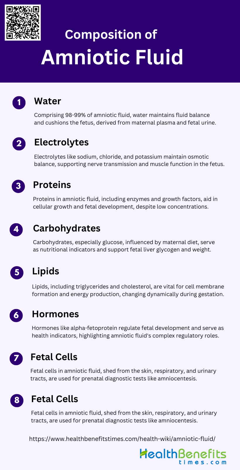
Amniotic fluid is a vital component in fetal development, providing protection and a stable environment for growth. Its composition is complex and includes various substances that support the fetus. The key components of amniotic fluid are:
1. Water
Amniotic fluid is primarily composed of water, which constitutes about 98-99% of its total volume. This water is crucial for maintaining the fluid balance and providing a protective cushion for the developing fetus. The water content in amniotic fluid is derived from both maternal plasma and fetal urine, ensuring a continuous exchange and circulation between the amniotic fluid and the fetal and maternal bloodstreams.
2. Electrolytes
Electrolytes such as sodium, chloride, and potassium are present in amniotic fluid and play a vital role in maintaining osmotic balance and proper cellular function. The levels of these electrolytes in amniotic fluid are similar to those found in maternal and fetal blood, indicating a dynamic equilibrium between these compartments. This balance is essential for the physiological functions of the fetus, including nerve transmission and muscle contraction.
3. Proteins
Proteins in amniotic fluid are significantly lower in concentration compared to maternal and fetal blood. These proteins include enzymes, growth factors, and other bioactive molecules that contribute to fetal development. The presence of specific proteins and proteases in amniotic fluid suggests roles in cellular growth, proliferation, and morphogenesis, highlighting the fluid’s importance beyond mere protection and waste regulation.
4. Carbohydrates
Carbohydrates, particularly glucose, are present in amniotic fluid and are influenced by maternal diet. Studies have shown that higher levels of maternal dietary carbohydrates can increase the glucose concentration in amniotic fluid, which is positively associated with fetal liver glycogen and fetal weight. This indicates that amniotic fluid glucose levels can serve as a nutritional indicator for the developing fetus.
5. Lipids
Lipids in amniotic fluid are present in lower concentrations compared to maternal and fetal blood. These lipids, including triglycerides and cholesterol, are essential for fetal development, particularly for the formation of cell membranes and the production of energy. The dynamic changes in lipid levels throughout gestation suggest their involvement in various stages of fetal growth and development.
6. Hormones
Hormones in amniotic fluid, such as alpha-fetoprotein and other tumor markers, display different dynamic changes compared to maternal and fetal blood. These hormones play crucial roles in regulating fetal development and can serve as indicators of fetal health and development. The presence of these bioactive molecules underscores the complex regulatory functions of amniotic fluid.
7. Fetal Cells
Amniotic fluid contains fetal cells, which are shed from the fetal skin, respiratory tract, and urinary tract. These cells can be used for prenatal diagnostic tests, such as amniocentesis, to detect genetic abnormalities and fetal health conditions. The presence of fetal cells in amniotic fluid provides valuable information about the genetic and developmental status of the fetus.
8. Waste Products
Waste products in amniotic fluid, including urea, uric acid, and creatinine, are primarily derived from fetal urine. These waste products are indicative of fetal kidney function and metabolic activity. The levels of these substances can provide insights into the metabolic status of the fetus and are essential for monitoring fetal health and development.
Functions of Amniotic Fluid
Amniotic fluid plays a crucial role in the development and protection of the fetus throughout pregnancy. It provides a supportive environment that facilitates various physiological processes essential for fetal growth. The primary functions of amniotic fluid include:
1. Cushioning and Protection
Amniotic fluid (AF) plays a crucial role in cushioning and protecting the fetus from external shocks and mechanical injuries. The fluid creates a buoyant environment that allows the fetus to move freely, which helps in distributing any external pressure evenly across the fetal body, thereby minimizing the risk of injury. This protective function is essential for the overall well-being of the fetus, as it ensures a stable and secure environment for growth and development.
2. Temperature Regulation
Amniotic fluid is vital for maintaining a consistent temperature around the fetus, which is crucial for its development. The fluid acts as a thermal buffer, absorbing and dissipating heat to ensure that the fetus remains at an optimal temperature. This thermoregulatory function is particularly important because fluctuations in temperature can adversely affect fetal development and lead to complications.
3. Infection Prevention
The amniotic fluid contains various antimicrobial peptides, cytokines, and immune cells that contribute to its role in preventing infections. These components of the innate immune system help protect the fetus from microbial invasions that could lead to intra-amniotic infections and inflammation (IAI). The presence of these immune factors in the amniotic fluid is crucial for safeguarding the fetus against potential pathogens.
4. Lung and Digestive System Development
Amniotic fluid is essential for the development of the fetal lungs and digestive system. The fetus breathes in and swallows the fluid, which helps in the maturation of the lungs and the gastrointestinal tract. This process prevents pulmonary hypoplasia and promotes the development of the digestive system by exercising the muscles involved in swallowing and digestion.
5. Movement and Muscle Development
The presence of amniotic fluid allows the fetus to move freely within the womb, which is crucial for the development of muscles and bones. This movement helps in the proper formation and strengthening of the musculoskeletal system. The fluid provides a medium in which the fetus can practice movements, thereby aiding in muscle development and coordination.
6. Nutrient Exchange
Amniotic fluid serves as a medium for the exchange of nutrients between the mother and the fetus. It contains various nutrients, including proteins, carbohydrates, lipids, and electrolytes, which are essential for fetal growth and development. The fluid also facilitates the transfer of growth factors and other bioactive molecules that support cellular growth and proliferation.
Abnormalities and Their Implications of Amniotic fluid
Abnormalities in amniotic fluid can significantly impact fetal development and pregnancy outcomes. These abnormalities may involve either an excess or deficiency of the fluid, each carrying its own set of risks and complications. The main types of amniotic fluid abnormalities and their implications include:
1. Polyhydramnios
Polyhydramnios, characterized by an excessive volume of amniotic fluid, complicates 1-3% of pregnancies and is often associated with fetal abnormalities, maternal diabetes, and twin pregnancies, though 60% of cases are idiopathic. This condition is linked to higher perinatal mortality rates (10-30%) and an increased risk of preterm birth (up to 22%). Fetal anomalies such as esophageal and intestinal atresias, neural tube defects, and central nervous system malformations are commonly associated with polyhydramnios. Additionally, pregnancies complicated by polyhydramnios often result in higher birth weights, increased cesarean section rates, and a greater frequency of maternal morbidity, including conditions like hypertension and diabetes mellitus. The neonatal outcome largely depends on the underlying pathology, with gastrointestinal tract abnormalities showing relatively good prognoses post-surgery.
2. Oligohydramnios
Oligohydramnios, defined by a decreased volume of amniotic fluid, affects 3-5% of pregnancies and is frequently caused by premature rupture of membranes, fetal urinary tract malformations, chromosomal abnormalities, and certain medications like NSAIDs. This condition is associated with lower birth weights, shorter gestational ages, and higher rates of spontaneous abortions. Severe oligohydramnios can lead to pulmonary hypoplasia, significantly impacting neonatal survival, although amnioinfusion can sometimes improve outcomes by restoring amniotic fluid volume. The condition is also linked to increased rates of cesarean sections, neonatal intensive care unit admissions, and neonatal mortality. Proteomic studies suggest that idiopathic oligohydramnios may involve alterations in cellular organization and immunological functions, as well as increased activity in vesicular transport pathways across the amnion.
Diagnostic Tests of Amniotic fluid
Diagnostic tests of amniotic fluid are essential for assessing fetal health and detecting potential abnormalities during pregnancy. These tests provide valuable information about genetic conditions, infections, and the overall well-being of the fetus. The primary diagnostic tests of amniotic fluid include:
1. Amniocentesis
This procedure is usually conducted from the 16th week of pregnancy onwards to obtain fetal cells and other substances for various chromosomal, biochemical, molecular, and microbial studies. The primary reasons for performing amniocentesis include prenatal diagnosis of chromosomal abnormalities, single gene disorders, fetal infections, and intra-amniotic inflammation. It is also used to assess fetal lung maturity and blood or platelet type. Despite its diagnostic benefits, amniocentesis carries a risk of fetal loss (approximately 0.5%) and other complications such as amniotic fluid leakage and placental hemorrhage.
2. Ultrasound
Ultrasound is a non-invasive imaging technique widely used in prenatal care to assess the amniotic fluid volume and monitor fetal development. High-resolution ultrasound is particularly valuable in managing pregnancies at risk for conditions like oligohydramnios, which is characterized by low amniotic fluid levels. This technique helps in the early diagnosis of fetal abnormalities and guides further evaluation and management. Ultrasound is also essential in later stages of pregnancy to assess fetal lung maturity and detect potential complications such as Rhesus immunization. The accuracy and non-invasive nature of ultrasound make it a crucial tool in prenatal diagnostics.
3. Non-Stress Tests
Non-stress tests (NST) are a common method of antenatal fetal assessment introduced in the 1970s. This test monitors fetal heart rate in response to fetal movements, providing insights into fetal well-being without inducing stress. NST is particularly useful for high-risk pregnancies where other tests like the contraction stress test may be contraindicated. The test is often combined with other assessments such as vibroacoustic stimulation and biophysical profiles to provide a comprehensive evaluation of fetal health. Despite its widespread use, further research is needed to optimize its application and improve predictive accuracy.


