 The part of the body with which a person sees.
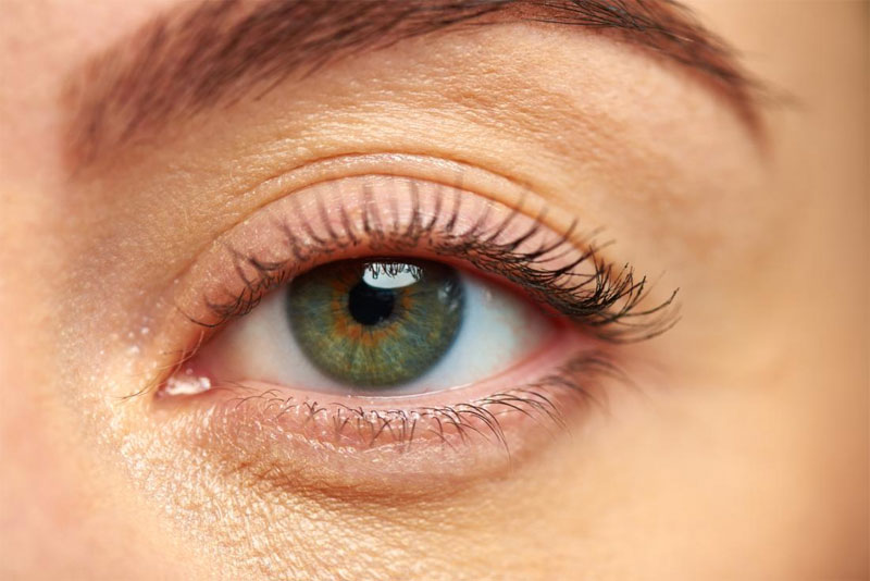
Either of two organs of sight located in a bony socket at the front of the skull. The outer coat of the eye is made of the anterior transparent cornea and the posterior opaque sclera (white of the eye). Light enters through the cornea, passes through a fluid-filled anterior chamber, then passes through the opening (pupil) in the iris (colored portion of the eye) and finally through the lens and vitreous humor to strike nerve cells (rods and cones) in the retina of the eye. The impulse is then transmitted form the retina through the optic nerve to the brain.
The sensory organ of sight. The eyes focus light rays to perceive images and then convert these images into impulses that are transmitted to the brain.
The organ of sight: a three-layered roughly spherical structure specialized for receiving and responding to light.
The eye is the sensory organ of sight. It is an elaborate photoreceptor detecting information, in the form of light, from the environment and transmitting this information by a series of electrochemical changes to the brain. The visual cortex is the part of the brain that processes this information (i.e. the visual cortex is what ‘sees’ the environment). The eyes sit within bony cavities (orbits). Each orbit is situated on the front of the skull, one on each side of the nose. The eye consists of an outer wall of three main layers, and a central cavity divided into three.
The sense organ that gathers and focuses light, generates signals that are sent to the brain, and
allows one to see.
The eyes function together, coordinated by the brain, to focus on a specific object and create a clear image on each retina. To achieve this, the eyes align themselves appropriately. When needed, they can enhance the image’s clarity by adjusting the focus through an automatic process known as accommodation, which involves changing the shape of the lens.
The eyeballs are situated within fatty pads inside the bony orbits, or eye sockets, which offer protection against injuries. To facilitate movement, each eyeball is controlled by six delicate muscles. The eye has a durable, white outer layer called the sclera. Positioned at the front of the sclera, the cornea is a transparent, thin-walled dome that acts as the primary “lens” of the eye, responsible for most of the focusing process.
Located behind the cornea, there is a shallow chamber that contains watery fluid known as the aqueous humor. Towards the back of this chamber lies the iris, which is the colored part of the eye, surrounding the pupil—a circular opening that appears black. The size of the pupil can be changed by tiny muscles in response to variations in light intensity. This mechanism allows the eye to control the amount of light entering, regulating vision accordingly.
Behind the iris and in direct contact with it, you can find the crystalline lens. This lens is suspended by fibers from a circular ring of muscle known as the ciliary body. When the ciliary body contracts, it alters the shape of the lens, allowing for precise fine-tuning of focus.
The primary inner part of the eye contains a transparent gel called the vitreous humor and is situated right behind the lens. At the back interior of the eye is the retina, which is a complex structure made of nerve tissue and highly sensitive to light. The retina constantly needs a supply of oxygen and glucose to function properly. To fulfill this requirement, there is a thin network of blood vessels known as the choroid plexus located directly beneath it. The choroid is connected to the ciliary body and the iris at the front, and together, these three parts make up the uveal tract.
The eyeball is enclosed by a transparent and flexible membrane known as the conjunctiva. This membrane is attached to the skin at the corners of the eye and forms the inner lining of the eyelids. Within the conjunctiva, there are glands that secrete mucus. Along with the meibomian glands found in the eyelids, which secrete an oily fluid, they work together to create the tear film that serves to protect the cornea and conjunctiva of the eye. When we blink, it is a protective reflex that helps evenly spread the tear film over the cornea, ensuring clear vision and providing essential eye protection.
The organ of sight. It consists of a tough, dense membrane forming a globe, the front translucent portion of which is called the cornea. The front of the globe is covered by mucous membrane, which also lines the lids, called conjunctiva, and when this becomes inflamed the eye becomes red and irritable the condition is called conjunctivitis. Inside the eye is the crystalline lens, which can have its shape altered by muscles in order to focus clearly. When the lens becomes opaque it is called a cataract. In middle life the lens becomes stiff and inelastic and thus cannot be focused so well on near objects. The condition is called presbyopia and can be recognized by the need to wear reading spectacles. The chambers of the eye both in front of and behind the lens are filled with a substance called the aqueous humor. At the back of the eye the optic nerve enters from the brain and expands to form a visual sensitive plate, the retina, which has a small blind spot where the nerve enters it. The retina can be examined by an instrument called an ophthalmoscope. This blind spot is important to doctors because it is the one spot where veins and arteries can be examined under working conditions and because this area shows the early signs of pressure within the skull.